
Digital dentistry refers to the technologies, methodologies and devices that incorporate digital or computer-aided workflow instead of using electrical or mechanical tools. Carrying out dental processes can be much more efficient and precise whether we are talking about diagnostics, restorations or even surgery.
Digital dentistry technologies:
- CAD/CAM
- 3D printing
- Intraoral cameras and scanners (for digital impressions)
- Digital X-ray and CT
- Digital photos
- Digital smile design
- Guided surgery
- Digitally planned dental implant location: allows for dental implantation at places only available for implantation if preceded by bone regeneration.
- Guided surgery: Placing implants through a surgical guide has a lot better accuracy than with free hand. Can be done without flap preparation or stiches, what is more it can be performed with a minimal invasive approach without postoperative pain and swelling
- Digital smile design: your individual smile is planned before the actual treatment.
- Digital impressions: maximum precision for tooth replacement without impression material and discomfort
- Digitally planned and manufactured tooth replacements: CAD/CAM technology for every tooth replacement for maximal precision
- The perfect Hollywood smile: digitalizing workflows for the perfect result
CAD/CAM technology
Numerous studies have shown that digital design and manufacturing is much more precise than traditional technologies. Digital design incorporates CAD/CAM (computer-aided Designing/Computer Assisted Manufacturing) technology that has fundamentally changed dentistry in recent decades. Digital transformation is becoming increasingly important in our profession.
Our dental technicians have been using CAD/CAM technologies for over 20 years in order to produce zirconium, cobalt-chromium, titanium and lithium-disilicate work. The impressions and samples are scanned in the laboratory and the replacements are digitally designed and manufactured (most frequently used technologies are lathing or 3D printing)
Before digital dentistry we used conventional impressions, which meant several sources of error (saliva, blood, deformation of the material, sensitivity to heat and moisture, defects in the impression itself and the whole procedure taking too long etc.) The impression was then sent to the dental laboratory (here we would like to pinpoint that there were sometimes problems with the transport of the impressions; they got lost, went damaged, and broken or the driver had an accident) where a sample was produced with a special plaster that was also source further source of error. Only after that could we begin with actual production.
This procedure is now digitalized, meaning that we take a digital impression at first and this will then be manufactured digitally.
Scans
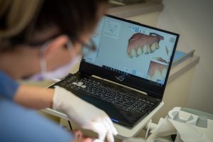
Scans are made with an intraoral scanner, without any traditional impression material or tray. This creates a 3D copy of the teeth that can be viewed on the computer immediately. One of the advantages of scans is that they do not cause any inconvenience for the patients; we can avoid the disturbing qualities of the impression material. The accuracy of scans can be measured in micrometres; therefore, they are extremely accurate.
The intraoral scanner used in our dental clinic can reproduce not only the shape, but the colours as well. Our patient can view the scan can together with the dentist. When using conventional impressions errors and inaccuracies are revealed only when the plaster form is ready. In this case the patient is usually called back for another appointment and we need to start the procedure over. A digital scan eliminates all these errors. Additionally, they are environmentally friendly due to the fact that we can reduce the use of materials, thus producing less waste.
Digital smile design
Considering the facial character of our patient we can create the smile that best suits him/her. We also take into account the shape and symmetry of the face, age and current smile in order to produce/create the temporary or permanent tooth replacements (dental crowns, bridges, dentures), restorations (e.g., veneers, whether direct or indirect) to be the best fit to the individual.
This can be done in several ways; the easiest way is to create a “visual design” based on photos. A more more modern solution is if we do the whole design in 3D by using a scanner and digital design. In this was the design process is completely interactive, we design your smile according to your individual needs!
Digital X-Ray, CT (CBCT)
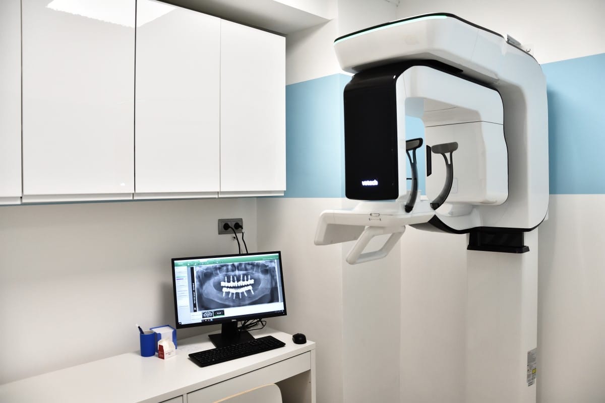
With digital X-Rays we use a special sensor that is placed in the mouth that is covered in a disposable pouch. Thanks to this, the scan is completely digital, the radiation exposure is much smaller and we get a well-detailed image.
During a cone beam CT examination, the C-arm or gantry rotates around the head in a complete 360-degree rotation while capturing multiple images from different angles that are reconstructed to create a single 3D image.
All data retrieved by the software is saved as virtual anatomy on the computer. The dentist can create different 3D views, which we call multiplanar reconstructions (MPR- multiplanar reconstructions).
We can choose between the different views and adjust the thickness of the slices based on the information we would like to visualize. The slices can be set in any direction and thickness to evaluate properly the critical area.
In addition, the CBCT has a three-dimensional view, what we can rotate to analyse it from any direction. One of the biggest advantages that the dose of the radiation it is just a fragment of a conventional CT (the modern CBCT scanner’s radiation dose is close to a panoramic X-ray, which is approximately equivalent to a 5-hour long flight). Thanks to the software, we can also convert the three-dimensional image into conventional radiological images.
Thanks to the almost endless possibilities offered by the software, the following can be diagnosed:
- Diseases, pathological irregularities
- Evaluating the possibility of a dental implant (bone volume and quality)
- The exact shape and position of the wisdom teeth.
- The course of nerve channels
- Sinus morphology
- Caries
- Lesions around roots
- Paradental problems
- Fractured teeth
- Jawbone joint and its diseases
- Bone lesions
- Bone fractures
Guided surgery
Simply put, based on a special dental CT scan (computer tomography), we can plan exactly where we would like to insert the implants or where they would be in the most optimal position. Based on this scan, we digitally design the surgical guide.
The surgical guide then is made by milling or 3D printing. After meticulous disinfection we secure the surgical guide on the jaw and we prepare the implant beds through the guide. The implants also can be inserted through the surgical template. Thanks to special sleeves incorporated in the guide the lateral movements of the drills are not permitted, so the accuracy of positioning the implants is a lot better than with the free-handed approach.
Arguments for guided surgery
Benefits for the patients
- Safety
- The implants placed at the optimal positions at safe distance from anatomical structures such as nerves or the paranasal sinus
- Raising a flap is not mandatory, the implants can be placed with a minimal invasive approach
- Comfort
- The overall time of the operation is shorter
- Less discomfort (swelling) after the surgery
- Less pain, sometimes it can be totally painless afterwards
- Thanks to the optimal implant position temporary/immediate restorations can be made right after the surgery
- Cost efficiency
- The surgical guide might involve extra costs, yet certain bone regenerative treatments can be avoided that might be further additional costs
- Less check-ups are necessary, the whole course of the treatment can be reduced
- We can assess at planning already what kind of abutments and supplementary parts are necessary. The outcome can be fully foreseen.
- Precision
- The implants set more parallel, so the distribution of the forces is more beneficial.
- The more precisely we design and insert the implants, the higher quality restorations can be made of titanium or zirconia.
- Thanks to accurate planning we can anticipate less complications (chipping, fracture etc.) on the long run after the delivery of the definitive restorations.